Our Services
Optical Dispensing
Palozej Eyecare has recently expanded our Optical Dispensary at each of our locations. We now offer an even greater selection of the most fashionable and durable frames available. Our Optical staff will help with the selection of the best frame as well as the best lenses for your needs. Please click below to visit some of our vendors for more information and selections!
Vendors
Comprehensive Eye Exam
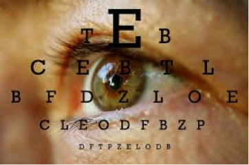
A Comprehensive Eye Exam is a complete evaluation of the eye as well as a full review of patient personal and family health history. The eye exam includes testing of Visual Acuity, Pupils, Eye Muscles, Stereopsis, Peripheral Vision, Color Vision, Refraction, Eye Pressure, Anterior Segment and Retina. The health history is a complete Review of Systems.

Contact Lenses
Palozej Eyecare carries all types of contact lenses. From common fitting of soft contacts for myopia (nearsighted) and hyperopia (farsighted) as well as more complicated fitting of presbyopia (bifocal) and astigmatism (weighted) we will work to ensure you have the best vision as well as the most comfortable contacts. We also do fittings for gas permeable or hard contacts for even more complicated cases such as keratoconus (irregular cornea).
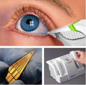
Digital Retinal Photography
Retinal (fundus) photography is used by your doctor to monitor the progression of eye diseases such as macular degeneration and glaucoma as well as systemic diseases such as diabetes. For more invitation, please visit Aetna.
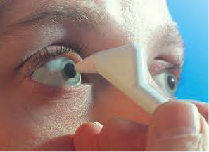
Dilated Eye Exam
During an eye exam, your doctor may use one of several different eye drops to help widen the black center of your eye (pupil). The strength of the eye drops may depend on the reason for your eye exam. Routine dilation of the pupil helps your doctor diagnose many eye diseases or conditions such as diabetes, high blood pressure and macular degeneration. Maximum dilation of the pupil may be needed to help your doctor diagnose such conditions as retinal detachment. For more information, please visit Mayo Clinic.
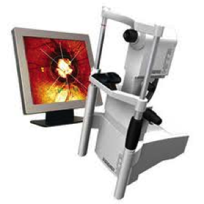
HRT
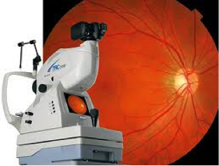
Heidelberg Retinal Tomograph measurements of the optic nerve and retinal nerve fiber layer (RNFL) are an increasingly important aspect of the early diagnosis and then treatment of glaucoma. Detecting micrometer changes has allowed doctors to make a diagnosis of glaucoma more accurately than ever before. For more information, please visit Heidelberg Engineering.
Laser Vision Correction Consult and Co-Management
Lasik (Laser-Assisted-In-Situ Keratomileusis) is the leading laser procedure for the correction of visual refractive errors. Today, nearly everyone is a candidate for the procedure. Although Optometrists can perform Lasik in other states, Optometrists in Connecticut currently Co-Manage. Please ask our doctors if you have any questions about refractive surgery. For more information, please visit TLC Laser Eye Associates.
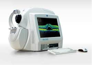
Laser Vision Correction Consult and Co-Management
Optical Coherence Tomography is an exciting new imaging technique that obtains micrometer resolution and 3D views of the retina. It is used to help your doctor diagnose and monitor treatment for conditions such as macular degeneration (both wet and dry), macular hole, macular edema, diabetic retinopathy and glaucoma. For more information, please visit Zeiss Cirrus HD-OCT.
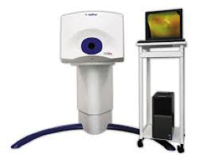
Optos Advanced Retinal Photography
Optos retinal imaging uses an ultra-widefield scanning laser to obtain a view of the retina that can be up to 6x wider than standard retinal photography. This allows your doctor to better diagnose and monitor conditions of the peripheral retina and choroid such as retinal detachment, retinoschisis, lattice degeneration, and paving stone degeneration. For more information, please visit Optos.
Pachymetry
Pachymetry is the measurement of the thickness of the central cornea. Since the release of the Ocular Hypertension Study (OHTS) in 2002, eye doctors have realized that corneal thickness plays a major role in glaucoma. For more information, please visit the Glaucoma Research Foundation.
Tear Lab Dry Eye Testing
Dry eye has become an increasingly common eye problem. The osmolarity (salt content) of tears has recently been shown to be the best test to diagnose and then monitor Dry Eye Disease. As a CLIA federal certified laboratory, our doctors can use the Tear Lab to better diagnose, treat and monitor dry eye disease. For more information, please visit the Tear Lab.
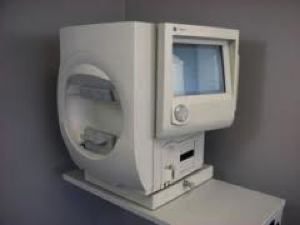
Visual Field Testing
Automated Perimetry is a thorough evaluation of your central and peripheral visual field. Eye diseases such as glaucoma and systemic diseases such as diabetes can damage your peripheral visual field. Brain abnormalities such as stroke or tumors can also affect your visual field. The location in the brain of a stroke or tumor can often be determined by the size, shape and location of a visual field defect. For more information, please visit the Glaucoma Research Foundation.